Human Leg Bone Diagram / Label the Bones of the Leg -- Exploring Nature Educational ... : Pelvic bone labeled 12 photos of the pelvic bone labeled pelvic bone labeled, pelvic bone labeling quiz.
Human Leg Bone Diagram / Label the Bones of the Leg -- Exploring Nature Educational ... : Pelvic bone labeled 12 photos of the pelvic bone labeled pelvic bone labeled, pelvic bone labeling quiz.. Continue scrolling to read more below. This diagram depicts leg parts anatomy with parts and labels. See more ideas about muscle anatomy, human anatomy and physiology, body anatomy. The foot bones shown in this diagram are the talus, navicular, cuneiform, cuboid, metatarsals and calcaneus. Become a pro with these valuable skills.
This diagram depicts leg parts anatomy.human anatomy diagrams show internal organs, cells, systems, conditions, symptoms and sickness information and/or tips for healthy living. Posted on june 8, 2016 by admin. This diagram depicts leg parts anatomy with parts and labels. The ilium is the big bone of the hip, the ischium is the bone on which one sits and the pubis forms the lower frontal hip bone as seen in the diagram. The human leg, in the general word sense, is the entire lower limb of the human body, including the foot, thigh and even the hip or gluteal region.
Appendages - Skeletal Learning from skeletallearning.weebly.com B a connects leg to hip match each of the facts below to the correct joint (write them in the correct column). Human leg osteoarthritis inflammation of bone joints. Pelvic bone labeled 12 photos of the pelvic bone labeled pelvic bone labeled, pelvic bone labeling quiz. This diagram depicts leg parts anatomy.human anatomy diagrams show internal organs, cells, systems, conditions, symptoms and sickness information and/or tips for healthy living. Beside that, we also come with more related ideas as follows free printable human anatomy coloring pages, lower leg muscle diagram blank and lower limb bones unlabeled. The bones together make up the hip. Posted by kevin lee on 8 april 2019, 10:24 am. Join over 50 million people learning online at udemy!
Structure of anatomy leg and foot 6 photos of the structure of anatomy leg and foot leg foot anatomy, leg foot bones, leg foot cramps, leg foot cramps at night, leg foot massage, leg foot numbness, leg foot pain, leg foot tattoos, foot, leg foot anatomy, leg foot.
The femur or the thigh bone is closest to the body. The bones of the leg are the femur, tibia, fibula and patella.the foot bones shown in this diagram are the talus, navicular, cuneiform, cuboid, metatarsals and calcaneus. The femur, or thigh bone, is the largest, heaviest, and strongest bone in the human body. The knee joint is the largest joint in the body and is primarily a hinge joint, although some sliding and rotation occur. The thigh bone, or femur, is the large upper leg bone that connects the lower leg bones (knee joint) to the pelvic bone (hip joint). Related posts of leg bones anatomy diagram structure of anatomy leg and foot. The bones of the superior portion of the skull are known as the cranium and protect the brain from damage. The human leg, in the general word sense, is the entire lower limb of the human body, including the foot, thigh and even the hip or gluteal region. Labeled human leg bones created for use in leg bone. Beside that, we also come with more related ideas as follows free printable human anatomy coloring pages, lower leg muscle diagram blank and lower limb bones unlabeled. Structure of anatomy leg and foot 6 photos of the structure of anatomy leg and foot leg foot anatomy, leg foot bones, leg foot cramps, leg foot cramps at night, leg foot massage, leg foot numbness, leg foot pain, leg foot tattoos, foot, leg foot anatomy, leg foot. The fibula is connected via ligaments. This diagram depicts leg parts anatomy.human anatomy diagrams show internal organs, cells, systems, conditions, symptoms and sickness information and/or tips for healthy living.
Related posts of leg bones anatomy diagram structure of anatomy leg and foot. The ilium is the big bone of the hip, the ischium is the bone on which one sits and the pubis forms the lower frontal hip bone as seen in the diagram. Join over 50 million people learning online at udemy! The longest and the strongest bone in the human skeletal system as you can observe in the labeled skeleton diagram of the human body. The femur or the thigh bone is closest to the body.
File:Human leg bones labeled.svg - Wikipedia from upload.wikimedia.org Pelvic bone labeled 12 photos of the pelvic bone labeled pelvic bone labeled, pelvic bone labeling quiz. The bones together make up the hip. This diagram depicts leg parts anatomy with parts and labels. For diagram showing its location relative to the fibula, tibia, patella, and other bones of the leg. Related posts of diagram of leg bones pelvic bone labeled. For more anatomy content please follow us and visit our website: The pubis, ischium, and ilium together constitute the pelvis while the thigh bone is the femur. The majority of muscles in the leg are considered long muscles, in that they stretch great distances.
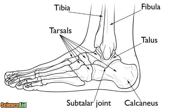
The foot bones shown in this diagram are the talus, navicular, cuneiform, cuboid, metatarsals and calcaneus.
The foot bones shown in this diagram are the talus, navicular, cuneiform, cuboid, metatarsals and calcaneus. Become a pro with these valuable skills. This image is an edited version of this image that was created by user:ladyofhats (mariana ruiz villarreal). The majority of muscles in the leg are considered long muscles, in that they stretch great distances. Human leg osteoarthritis inflammation of bone joints. These leg muscle diagrams show you the major muscles of the human leg. For more anatomy content please follow us and visit our website: B a connects leg to hip match each of the facts below to the correct joint (write them in the correct column). For diagram showing its location relative to the fibula, tibia, patella, and other bones of the leg. The femur, or thighbone, is the longest and largest bone in the human body. Also called the shin bone, the tibia is the longer of the two bones in the. Pelvic bone labeled 12 photos of the pelvic bone labeled pelvic bone labeled, pelvic bone labeling quiz. Join over 50 million people learning online at udemy!
The majority of muscles in the leg are considered long muscles, in that they stretch great distances. Human leg osteoarthritis inflammation of bone joints. Human legs, modeled at anatomynext.com, based on radiology scans, theime atlas of anatomy, and expert advise. Side view of foot bones inter mediate gone gone gone talus gone ca can eug gone cuboid gone gores phalange go neg o 5th metatarsal gone For more anatomy content please follow us and visit our website:
Bones of the Human Leg and Foot - ScienceAid from scienceaid.net The human leg, in the general word sense, is the entire lower limb of the human body, including the foot, thigh and even the hip or gluteal region. Continue scrolling to read more below. Human legs, modeled at anatomynext.com, based on radiology scans, theime atlas of anatomy, and expert advise. Posted on june 8, 2016 by admin. The bones of the leg are the femur, tibia, fibula and patella.the foot bones shown in this diagram are the talus, navicular, cuneiform, cuboid, metatarsals and calcaneus. At the same time, the bones and joints of the leg and foot must be strong enough to support the body's weight while remaining flexible enough for movement and balance. Leg bone anatomy diagram diagram of human leg human anatomy diagram 10 / 10 ( 1 vote ) in this image, you will find femur, medial epicondyle of the femur, patella, tibial tuberosity, anterior rest of the tibia, a medial surface of the tibia, lateral epicondyle of the femur, head of the fibula, fibula, medial malleolus of the tibia, lateral. The diagram shows a human leg bone and the joints at each end of it (a and b).
For more anatomy content please follow us and visit our website:
Structure of anatomy leg and foot 6 photos of the structure of anatomy leg and foot leg foot anatomy, leg foot bones, leg foot cramps, leg foot cramps at night, leg foot massage, leg foot numbness, leg foot pain, leg foot tattoos, foot, leg foot anatomy, leg foot. The lower leg is comprised of two bones, the tibia and the smaller fibula. Cross section human cartilage bone under microscope view. The majority of muscles in the leg are considered long muscles, in that they stretch great distances. The ilium is the big bone of the hip, the ischium is the bone on which one sits and the pubis forms the lower frontal hip bone as seen in the diagram. The human leg, in the general word sense, is the entire lower limb of the human body, including the foot, thigh and even the hip or gluteal region. Its lower end helps create the knee joint. The femur or the thigh bone is closest to the body. The foot bones shown in this diagram are the talus, navicular, cuneiform, cuboid, metatarsals and calcaneus. Human legs, modeled at anatomynext.com, based on radiology scans, theime atlas of anatomy, and expert advise. Human leg osteoarthritis inflammation bone joints. A hinge joint a ball and socket joint lets leg swing freely lets leg bend and straighten found in the knee joint b joint a each joint consists of four different tissues, choose the correct role of the. The bones of the leg are the femur, tibia, fibula and patella.the foot bones shown in this diagram are the talus, navicular, cuneiform, cuboid, metatarsals and calcaneus.
Its lower end helps create the knee joint leg bone diagram. B a connects leg to hip match each of the facts below to the correct joint (write them in the correct column).


0 Komentar